Low-field cardiovascular MRI
Whole-heart MRI at 0.55T
2023–2025
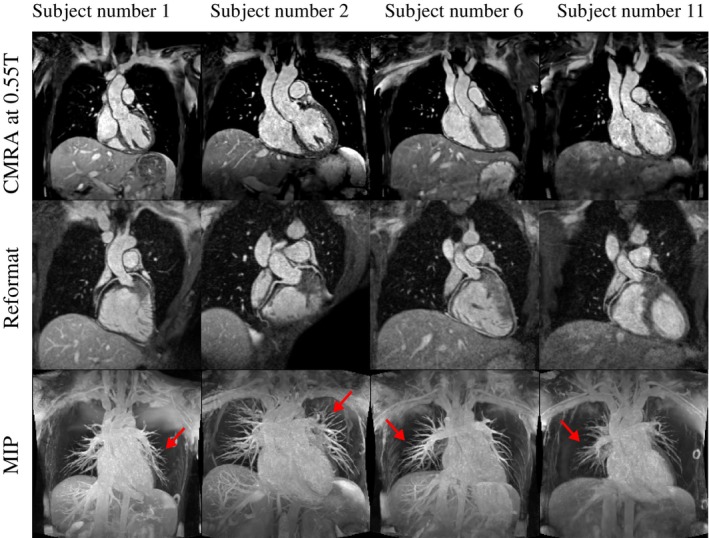
Free-breathing whole-heart MRI at 0.55T, combining coronary angiography with CMRA and simultaneous lumen and vessel-wall imaging with iT2prep-BOOST.
Why it matters
Low-field MRI can improve access to cardiovascular imaging, but reduced signal and different tissue properties require sequences to be reconsidered rather than transferred directly from higher field strengths.
Contribution
Both studies combine simulation-guided sequence design, image navigators, nonrigid respiratory motion correction, and patch-based denoising to support free-breathing whole-heart imaging at 0.55T.
Coronary angiography (CMRA)
The CMRA acquisition visualizes the main cardiac structures and coronary arteries without contrast agents. In the initial healthy-volunteer study, whole-heart images were acquired in approximately six minutes.
Lumen and vessel-wall imaging (iT2prep-BOOST)
iT2prep-BOOST produces co-registered 3D bright-blood and black-blood images in a single scan, providing complementary views of the vessel lumen and wall. The initial 0.55T study demonstrated this approach in healthy volunteers with a scan time of approximately eight minutes.
The subsequent MRM study extends iT2prep-BOOST to aortic imaging at either systole or diastole. It evaluates T2-preparation pulses to reduce flow-related artifacts, demonstrating simultaneous lumen and vessel-wall imaging in approximately seven minutes.
Resources
Project record
Publications and presentations
2025
Journal article
Simultaneous 3D aortic lumen and vessel wall imaging at 0.55 T at either systole or diastole
Magnetic Resonance in Medicine
2025
Journal article
Highly efficient image navigator based 3D whole-heart cardiac MRA at 0.55T
Magnetic Resonance in Medicine
Awards & recognition
- 2026 · Top Viewed Article in MRM (Top 10%)
2024
Oral presentation
Simultaneous iNAV-based 3D Whole-Heart Bright-Blood and Black-Blood Imaging at 0.55T
SCMR 2024 · London, United Kingdom
Awards & recognition
- 2024 · Early Career Award (1st Place), Basic Science, SCMR
2023
Oral presentation
Whole-Heart Cardiovascular Magnetic Resonance Angiography with iNAV-based Non-Rigid Motion-Corrected Reconstruction at 0.55T
ISMRM 2023 · Toronto, Canada
Awards & recognition
- 2023 · Summa Cum Laude Merit Award (Top 5%), ISMRM
- 2023 · MRM fast-track initiative